Cell and Tissue Research 404, 17. 2026.
Nueva publicación: Anatomy of the hypothalamic–pituitary axis and buccal lobe in the holocephalan Callorhinchus callorynchus
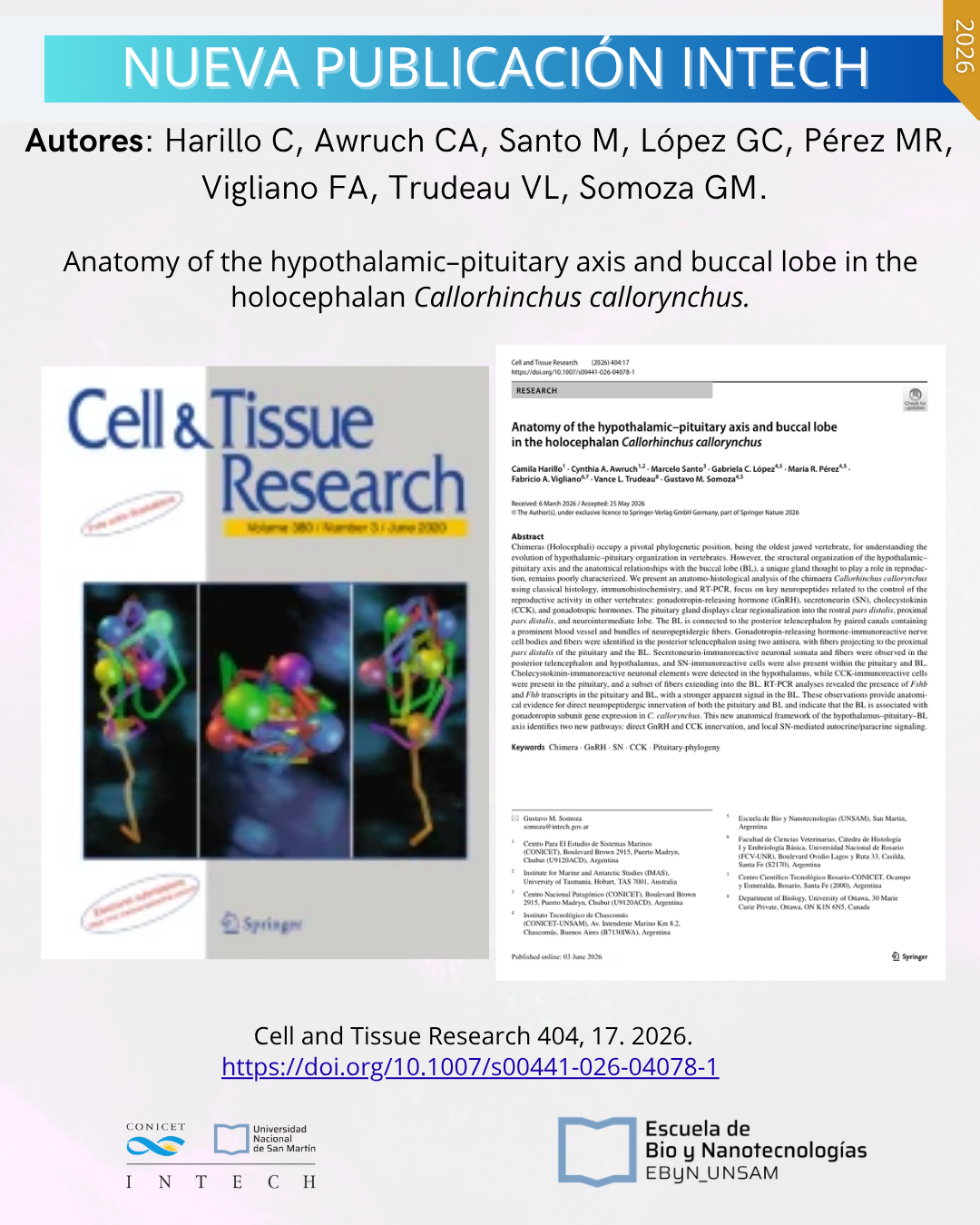
Chimeras (Holocephali) occupy a pivotal phylogenetic position, being the oldest jawed vertebrate, for understanding the evolution of hypothalamic–pituitary organization in vertebrates. However, the structural organization of the hypothalamic–pituitary axis and the anatomical relationships with the buccal lobe (BL), a unique gland thought to play a role in reproduction, remains poorly characterized. We present an anatomo-histological analysis of the chimaera Callorhinchus callorynchus using classical histology, immunohistochemistry, and RT-PCR, focus on key neuropeptides related to the control of the reproductive activity in other vertebrates: gonadotropin-releasing hormone (GnRH), secretoneurin (SN), cholecystokinin (CCK), and gonadotropic hormones. The pituitary gland displays clear regionalization into the rostral pars distalis, proximal pars distalis, and neurointermediate lobe. The BL is connected to the posterior telencephalon by paired canals containing a prominent blood vessel and bundles of neuropeptidergic fibers. Gonadotropin-releasing hormone-immunoreactive nerve cell bodies and fibers were identified in the posterior telencephalon using two antisera, with fibers projecting to the proximal pars distalis of the pituitary and the BL. Secretoneurin-immunoreactive neuronal somata and fibers were observed in the posterior telencephalon and hypothalamus, and SN-immunoreactive cells were also present within the pituitary and BL. Cholecystokinin-immunoreactive neuronal elements were detected in the hypothalamus, while CCK-immunoreactive cells were present in the pituitary, and a subset of fibers extending into the BL. RT-PCR analyses revealed the presence of Fshb and Fhb transcripts in the pituitary and BL, with a stronger apparent signal in the BL. These observations provide anatomical evidence for direct neuropeptidergic innervation of both the pituitary and BL and indicate that the BL is associated with gonadotropin subunit gene expression in C. callorynchus. This new anatomical framework of the hypothalamus–pituitary–BL axis identifies two new pathways: direct GnRH and CCK innervation, and local SN-mediated autocrine/paracrine signaling.
Harillo C, Awruch CA, Santo M, López GC, Pérez MR, Vigliano FA, Trudeau VL, Somoza GM. Anatomy of the hypothalamic–pituitary axis and buccal lobe in the holocephalan Callorhinchus callorynchus. Cell and Tissue Research 404, 17. 2026. https://doi.org/10.1007/s00441-026-04078-1